

Microscopy for Mushroom Identification
Use a microscope to examine spores, basidia, cystidia and tissues, the features needed to separate many difficult species.
Program identification
- 1.1Program name
- Microscopy for Mushroom Identification
- 1.2Type of qualification
- Course
- 1.3Provider / endorsing institution
- Fungaris – International Institute of Mycology, School of Culinary Mycology & Foraging. A Fungaris program of its own, not endorsed by a university.
- 1.4Mode of study
- Online, with live sessions in a small group, recordings and a virtual campus.
- 1.5Duration (hours / ECTS credits)
- 3 months (12 weeks) · 72 hours in total · 6 modules of 12 hours · 12 live sessions of 2 hours. ECTS credits: not applicable (Fungaris own program, not an official university degree).
- 1.6Language
- English
- 1.7Start and end dates / intake
- Next intake: January 11, 2027 to April 2, 2027. Enrollment closes on January 4, 2027. Following intake: March 8, 2027. How intakes work →
Overview
2.1Program description
This course opens up the microscopic features that separate many difficult mushroom species. Students learn how to choose a suitable microscope, set it up correctly and prepare clean mounts and sections. They then work with common reagents and stains, measure spores accurately, and examine the structures of the hymenium and cap cuticle, such as basidia, cystidia and pileipellis types. The final module shows how to feed microscopic data into technical keys to reach identifications that macroscopic features alone cannot support.
Many genera, from Inocybe to Russula and Cortinarius, cannot be reliably named without a microscope, and amateur mycologists increasingly contribute to serious recording. The program suits advanced foragers and amateur mycologists who already identify common species by eye. It is taught live by experienced mycologists who demonstrate techniques on screen. In the final project, each student produces a full microscopic description of a chosen species, with measured spores, annotated images and a key-based identification.
2.2Why it matters / what need it meets
It meets a practical need: applied, up-to-date knowledge in culinary mycology, useful in areas such as amateur mycology, biodiversity recording, natural history collections, ecological consultancy.
Mycology is moving fast and good practical training is still scarce. This program gives you a structured path, taught live by people who work in the field, so you can apply what you learn with judgment and safety.
Objectives and competences
3.1General objective
To finish the program able to apply its content professionally and with judgment. In short:
Use a microscope to examine spores, basidia, cystidia and tissues, the features needed to separate many difficult species.
3.2Specific objectives
- Build the foundations: Choosing and Setting Up a Microscope; Preparing Mounts and Sections.
- Put them into practice: Reagents and Stains; Measuring Spores.
- Apply everything to a real case: Hymenial and Cuticle Structures; Using Microscopic Data in Keys.
- Complete a final project reviewed by your instructor.
3.3Competences you will gain
- Set up and calibrate a microscope for fungal identification.
- Prepare clear mounts and sections from fresh and dried material.
- Use reagents such as Melzer's to test for diagnostic reactions.
- Measure spores accurately and report standard statistics.
- Apply microscopic characters in technical keys to confirm identifications.
Student profile
4.1Who it is for (entry profile)
Advanced foragers and amateur mycologists.
4.2Entry requirements
Good macroscopic identification skills are recommended. Students need access to a compound microscope with a 100x oil immersion objective.
4.3Career paths (exit profile)
- Amateur mycologist contributing to recording schemes
- Identification verifier for a mycological society
- Assistant in a herbarium or natural history collection
- Field survey assistant for ecological consultancies
- Pathway to advanced field mycology study
Outcomes depend on your experience and on your local market.
Study plan
5.1Overall structure (modules and workload)
| Module | Title | Phase | Topics | Hours |
|---|---|---|---|---|
| 01 | Choosing and Setting Up a Microscope | Foundations | 3 | 12 |
| 02 | Preparing Mounts and Sections | Foundations | 3 | 12 |
| 03 | Reagents and Stains | Practice | 3 | 12 |
| 04 | Measuring Spores | Practice | 3 | 12 |
| 05 | Hymenial and Cuticle Structures | Application & project | 3 | 12 |
| 06 | Using Microscopic Data in Keys | Application & project | 3 | 12 |
| Total | 72 |
6 modules × 12 hours = 72 hours: 24 hours of live sessions, and the rest for campus study, exercises and the final project.
5.2Content by module
Hover or tap a module to follow the path from the first session to the final project. Open a module to see the topics it covers.
- Compound microscope types and budgets
- Objectives, oil immersion and illumination
- Calibrating the eyepiece micrometer
- Squash mounts and spore preparations
- Cutting thin sections by hand
- Working with dried material
- Melzer's reagent and amyloid reactions
- Congo red and other stains
- Safe handling of reagents
- Measuring spore length and width
- Calculating Q values and averages
- Spore ornamentation and walls
- Basidia and their sterigmata
- Cheilocystidia and pleurocystidia
- Pileipellis and clamp connections
- Reading technical keys
- Combining macro and micro characters
- Writing a microscopic description
5.3Company placement
This program does not include a company placement. Practice is built into every module through exercises, case studies and the final project.
5.4Final project
A complete microscopic description of a chosen species, with measured spores, annotated images and a key-based identification.
Methodology
6.1How it is taught
- Live online sessions, usually weekly, with a working professional and a small group.
- Recordings of every session, so you can catch up if work or time zones get in the way.
- Exercises and real case studies in every module.
- Tutoring: your instructor answers questions and gives feedback on your exercises and project.
6.2Platform and resources
A Moodle virtual campus with session recordings, readings, exercises and forums for each module. You need a computer, an up-to-date browser and a stable connection; a webcam and microphone are recommended.
Technical requirements →6.3Estimated weekly workload
About 6 hours a week for 12 weeks: one 2-hour live session on a weekday evening, plus study and exercises on the campus at your own pace.
Assessment
7.1Assessment system and weighting
Progress is assessed through participation in live sessions, microscopy exercises in each module and a final project reviewed by the instructor.
7.2Requirements to pass
- Complete every module, by attending the live sessions or watching the recordings.
- Hand in the module exercises.
- Have your final project approved by your instructor.
Teaching team
8.1Academic direction
At Fungaris, the Culinary Mycology school coordinates the content and quality of this program. The academic director of each intake is named in the intake guide.
8.2Faculty
Taught by professionals who work in the field. The instructor for each intake is confirmed before the start.
Meet our faculty →Certificate
9.1Certificate awarded
Course Certificate
On completing the program and its project, Fungaris issues a Course Certificate. Fungaris certificates are the institute's own. Whether an employer, school or authority recognizes them depends on that organization and on local rules in each country.
9.2Issuing body / recognition
Issued by Fungaris (operated by Nexora Generation, S.L.) as a digital certificate. It is a Fungaris certificate of its own: it is not an official university degree and does not award ECTS credits.
About our certificates →Admission and enrollment
10.1Admission process (steps)
- Send your request through the form or write to admissions.
- Admissions reviews your profile and confirms the start date, the session schedule and the fee.
- You secure your place with full payment or a 40% reservation.
- You receive your campus access and welcome guide before the first session.
10.2Documents required
- A valid identity document.
- Contact and billing details (personal or company).
10.3Deadlines
Enrollment for the intake starting on January 11, 2027 is open until January 4, 2027, or until the group is full (maximum 20 students).
10.4Fee and payment options
Fee: $450
- Full payment before the start: $360 (20% reduction).
- 40% reservation ($180) and 3 monthly installments of $90.
10.5Grants, discounts and funding
Discounts: 10% for Fungaris alumni and 15% for groups of three or more people from the same organization. Discounts cannot be combined with each other or with the full-payment reduction.
Your employer can pay for the program and receive the invoice in the company name. Whether a public or employer training budget can cover the fee depends on your country, so check with them.
10.6Cancellation policy
The conditions for canceling, withdrawing or moving to a later intake are explained in our cancellation policy and in your enrollment confirmation. Nothing there limits your rights under the consumer law that applies to you.
Cancellation and refunds →Additional information
11.1Frequently asked questions
Do I need experience to study Microscopy for Mushroom Identification?
Good macroscopic identification skills are recommended. Students need access to a compound microscope with a 100x oil immersion objective.
How is Microscopy for Mushroom Identification taught?
Live online, in 6 modules with a working professional, a small group and a final project.
How am I assessed?
Progress is assessed through participation in live sessions, microscopy exercises in each module and a final project reviewed by the instructor.
What is the final project?
A complete microscopic description of a chosen species, with measured spores, annotated images and a key-based identification.
What certificate do I get?
On completing the program and its project, Fungaris issues a Course Certificate. Fungaris certificates are the institute's own. Whether an employer, school or authority recognizes them depends on that organization and on local rules in each country.
What can I study after Microscopy for Mushroom Identification?
The natural next step is Diploma in Culinary Mycology.
11.2Alumni testimonials
We only publish verified testimonials from real students. This program does not have any published yet; they will appear here once its first intakes finish.
11.3Admissions contact
Write to info@fungaris.institute or use the contact form. Admissions answers in your language.
Read before you start.

Keep branching.
From our species guide.
 EdibleCaesar's mushroomAmanita caesareaGilled mushrooms
 Deadly poisonousDeath capAmanita phalloidesGilled mushrooms
 Deadly poisonousDestroying angelAmanita virosaGilled mushrooms
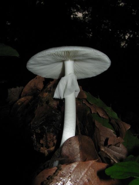 Deadly poisonousFool's mushroomAmanita vernaGilled mushrooms
 PoisonousGreen-spored parasolChlorophyllum molybditesGilled mushrooms
 PoisonousPanther capAmanita pantherinaGilled mushrooms
 EdibleSt George's mushroomCalocybe gambosaGilled mushrooms
 PoisonousYellow stainerAgaricus xanthodermusGilled mushrooms
Join the next intake.
Pay in full with a 20% reduction, or reserve your place with 40% and pay the rest in monthly installments. All prices are stated in US dollars (USD).